Breast Ultrasound Vs Mammogram: Which Is Right For You?
Find out more
Breast cancer is one of the most common types of cancer in the UK. Around 55,000 women in the UK are diagnosed with breast cancer every year. Your risk increases with age, so it’s important for women 40 and older to get routinely screened for breast cancer.
Breast cancer tests include mammograms and breast ultrasounds. Mammograms are considered the gold standard for breast cancer screening, but breast ultrasounds can be highly beneficial for some women.
This article will explore the technology of two diagnostic tests, their key advantages and disadvantages and what considerations you should be aware of when deciding if either a mammogram or breast ultrasound is right for you.
What is a breast ultrasound?
A breast ultrasound scan uses high-frequency sound waves to create computer images of the inside of the breast.
Breast ultrasounds are not typically done as part of regular breast cancer screening programmes, but they can be a vital diagnostic tool in some cases:
- For women with dense breasts (often younger women)
- When a lump can be felt but is not detected on a mammogram
- To differentiate between benign fluid-filled cysts and solid masses
During a breast ultrasound, the operator slides a handheld, wand-like instrument called a probe across your skin. The probe emits sound waves that pick up echoes as the waves bounce off your body tissues to create an image of the inside of your breast.
Breast ultrasounds are non-invasive and widely available.
What is a mammogram?
A breast screening mammogram is an X-ray examination of the breasts that uses low doses of radiation to detect cancer. Mammography can be used either for screening purposes (for early detection of breast cancer) or to make a diagnosis.
Women older than 30 years old should have a mammogram if they experience any symptoms of breast cancer, including:
- Breast pain
- Nipple discharge
- Palpable lumps
- Skin changes to the breast or nipple
- Any other noticeable changes to their breast size and shape
The NHS breast cancer screening programme consists of four mammograms (two for each breast). Women between the ages of 51 and 70 will be invited for a screening every three years.
Should you have a breast ultrasound or a mammogram?
Both breast ultrasounds and mammograms can be beneficial imaging tests for diagnosing breast cancer. Often, the two can complement each other, so it’s not a simple matter of choosing one or the other.
Each imaging technique has its benefits and limitations, which we’ll explore in more detail below. Ultimately, the most suitable test for you will depend on several factors, including your age, family history, symptoms and the recommendation of your GP.
Benefits
A mammogram is highly effective in detecting breast cancer early, when it is too small to see or feel. Mammograms look at changes in the breasts, and the radiologists who read your results will compare them against previous mammograms if you have any.
When looking at a mammogram, the doctor will be checking for breast changes such as:
- Calcification – The build-up of calcium within the breast tissue may be an early indication of breast cancer.
- Masses – Abnormal areas called masses can include cysts or solid masses that require further diagnostic testing.
- Asymmetries – Areas that look different from normal breast tissue patterns require additional diagnostic imaging to rule out cancer.
- Breast density – A measure of how much fibrous and glandular tissue is in your breast is called breast density. Women with higher breast density have a slightly higher risk of breast cancer. Additionally, the density makes it more difficult to spot abnormalities and cancer.
Breast ultrasounds can be helpful for women with dense breasts. In women younger than 40, the breast tissue is naturally denser (less fat), which makes it difficult for mammograms to detect change. Although typically a woman’s breasts become fattier as she ages, some women have dense breasts even as they get older. For this reason, women who are younger than 35 but who have a high risk of breast cancer due to their family history might be recommended annual breast ultrasound screenings.
The biggest advantage of breast ultrasounds is their use in diagnosing palpable breast lumps, especially those that can be felt but not seen on a mammogram. Breast ultrasounds use soundwaves to tell the difference between fluid-filled cysts (which are harmless) and solid masses, which might also be benign but require more testing.
Limitations
Neither mammograms nor breast ultrasounds are 100% accurate in detecting breast cancer. Both are subject to user error in the operator giving the exam and the radiologist who reads the results.
The biggest limitation of mammograms is for women with dense breasts. Furthermore, mammograms cannot distinguish between a fluid-filled cyst and a solid mass.
A breast ultrasound is conducted using a hand-held probe that slides along the skin. This means it has several inherent limitations, including:
- It does not provide a view of the entire breast
- It cannot examine abnormalities deep in the breast tissue
- It does not examine the axillary lymph nodes in the armpits
Risks and side effects
Although mammograms are generally considered safe, they do use small amounts of radiation. The amount of radiation is likely negligible for most women, but it’s not safe for pregnant women as it may harm the foetus.
As with most screening tests, there is also a risk of getting a false positive. This is when the mammogram picks up something that is not cancer. A false positive can cause anxiety and may lead to further tests such as a breast biopsy.
There are no side effects of breast ultrasounds. Because there is no risk of radiation, they are considered suitable for pregnant women.
Considerations
Anyone registered as female with their GP is eligible for the NHS national breast cancer screening programme. However, some considerations affect a woman’s decision about when and how often to get a mammogram.
Mammogram machines have two plates that compress or flatten the breasts to spread the breast tissue apart. It can be uncomfortable, so it’s recommended to schedule appointments the week after your period when your breasts are the least tender.
Mammograms for women with breast implants should still be done regularly, but both silicone and saline implants can make it more challenging to get high-quality images of the breast tissue. If you have breast implants, it’s essential to notify the clinic when making your appointment so they can arrange a technician who has experience in giving mammograms to women with breast implants.
Breast ultrasounds are simple, non-invasive and widely available. Their key strength is their ability to help diagnose palpable lumps. Despite this, it’s important to note that they are not suitable replacements for mammograms in the early detection of breast cancer. Breast ultrasounds are unable to detect calcification in the breast or provide a complete picture of the entire breast.
Breast ultrasound vs mammogram: which is best?
Despite the small amount of radiation and potential discomfort of testing, the benefits of mammograms far outweigh their limitations. Mammograms are highly effective in detecting abnormalities, masses and calcifications — all of which may be early indicators of breast cancer.
Breast ultrasounds, on the other hand, can be a very useful diagnostic imaging test, especially if a lump has been detected. They use sound waves and not radiation to create an image of the inside of your breasts, which makes them safer for pregnant women. However, the downside is that they are not able to capture images of the entire breast at once, reach the areas deep inside the breast or detect any calcifications.
When it comes to screening for breast cancer, a mammogram is considered the gold standard in detecting breast cancer.
Why should you book a breast screening mammogram?
Mammograms can be highly useful in detecting breast cancer in its early stages before any symptoms have been detected by you or your doctor. Women who are diagnosed with breast cancer following a routine screening usually require less treatment and have a better survival rate than those who present with symptoms.
Several extensive studies around the world show that breast cancer screening with mammograms reduces the number of deaths from breast cancer in women aged between 40-69, especially in those aged 50+.
Check4Cancer offers rapid access appointments for private breast screening mammograms to detect breast cancer at an early stage before changes can be felt in the breast. You don’t need a GP referral, and reports are given by highly experienced breast radiologists within three to four weeks.

Professor Simon Russell
Deputy Chief Medical Officer and Advisor for Breast Cancer
Consultant Oncologist at Addenbrooke’s Hospital, Cambridge and Hinchingbrooke Hospital, Huntingdon, Cambridgeshire. Professor of Oncology, University of Rome Medical School. Professor Simon Russell is a Consultant Oncologist at Addenbrooke’s Hospital, Cambridge and Hinchingbrooke Hospital, Huntingdon. He leads the Urological Malignancy Service and has previously been lead clinician for the breast service. He is the lead for Radiotherapy for Genesis Cancer Care, Cambridge. He has served as the secretary of the British Uro-Oncology Group 2003-2015 and remains a Trustee. He was appointed Professor of Oncology at the University of Rome Medical School, Tor Vergata 2015. Simon joined Check4Cancer in 2018.
Find out moreKnowledge and support
Go to all articles
What is Breast Cancer Awareness Month? All You Need to Know
First introduced in 1985, Breast Cancer Awareness Month runs every October with the initial aim of encouraging breast cancer screening...
Read more
Do Mammograms Hurt?
How a mammogram works, how long it takes, if it’s normal to hurt, and what to expect after a mammogram.
Read more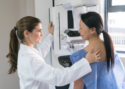
What Happens At A Mammogram?
Looking at what a mammogram detects, how often you should have a breast screening, and what happens before, during and...
Read more